What is Interlaminar Endoscopic Spine Surgery
Interlaminar Endoscopic Spine Surgery is a modern, minimally invasive technique used to treat various spinal conditions with less pain, faster recovery, and minimal tissue disruption. Through a small incision in the natural interlaminar window of the spine, surgeons use a high-definition endoscope to directly visualize and treat the affected nerves, discs, or ligaments.
This approach preserves normal anatomy, minimizes muscle damage, and offers quicker return to daily activities compared to traditional open surgery.
Types of Interlaminar Endoscopic Procedures
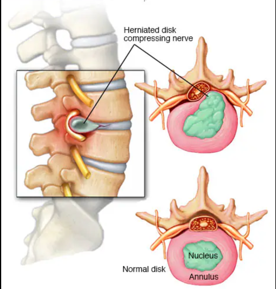
🔹 Interlaminar Endoscopic Discectomy
Removal of herniated disc material pressing on nerves. Ideal for central and paracentral herniations.
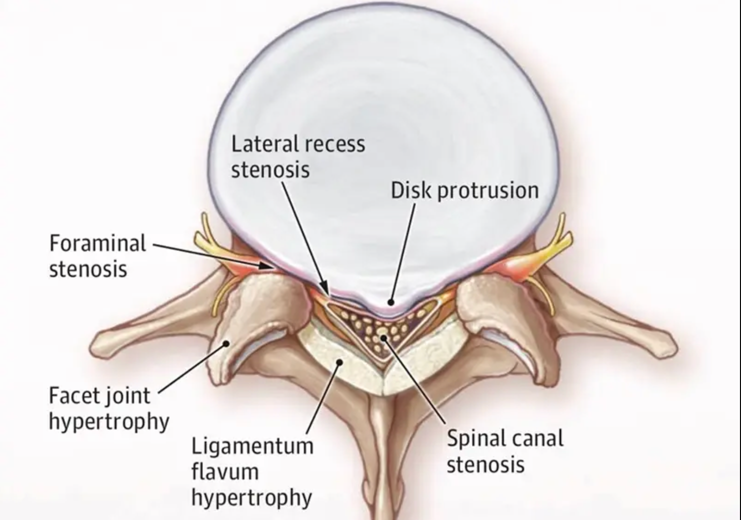
🔹 Interlaminar Endoscopic Decompression
Reduces nerve compression by removing thickened ligament or small bone spurs (commonly for lumbar canal stenosis).
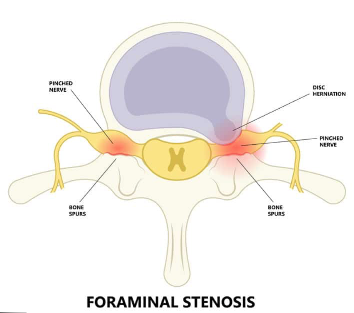
🔹 Endoscopic Foraminotomy (Interlaminar Route)
Enlarges the spinal nerve exit canal to relieve radiating leg pain from foraminal stenosis
🔹 Interlaminar Endoscopic Decompression (LE-ULBD)
LE-ULBD (Lumbar Endoscopic Unilateral Laminotomy for Bilateral Decompression) is a minimally invasive endoscopic technique used to treat lumbar canal stenosis.
It removes thickened ligament, hypertrophied bone, and soft tissue causing narrowing — using a unilateral approach to decompress both sides of the spinal canal while preserving stability..
Indications
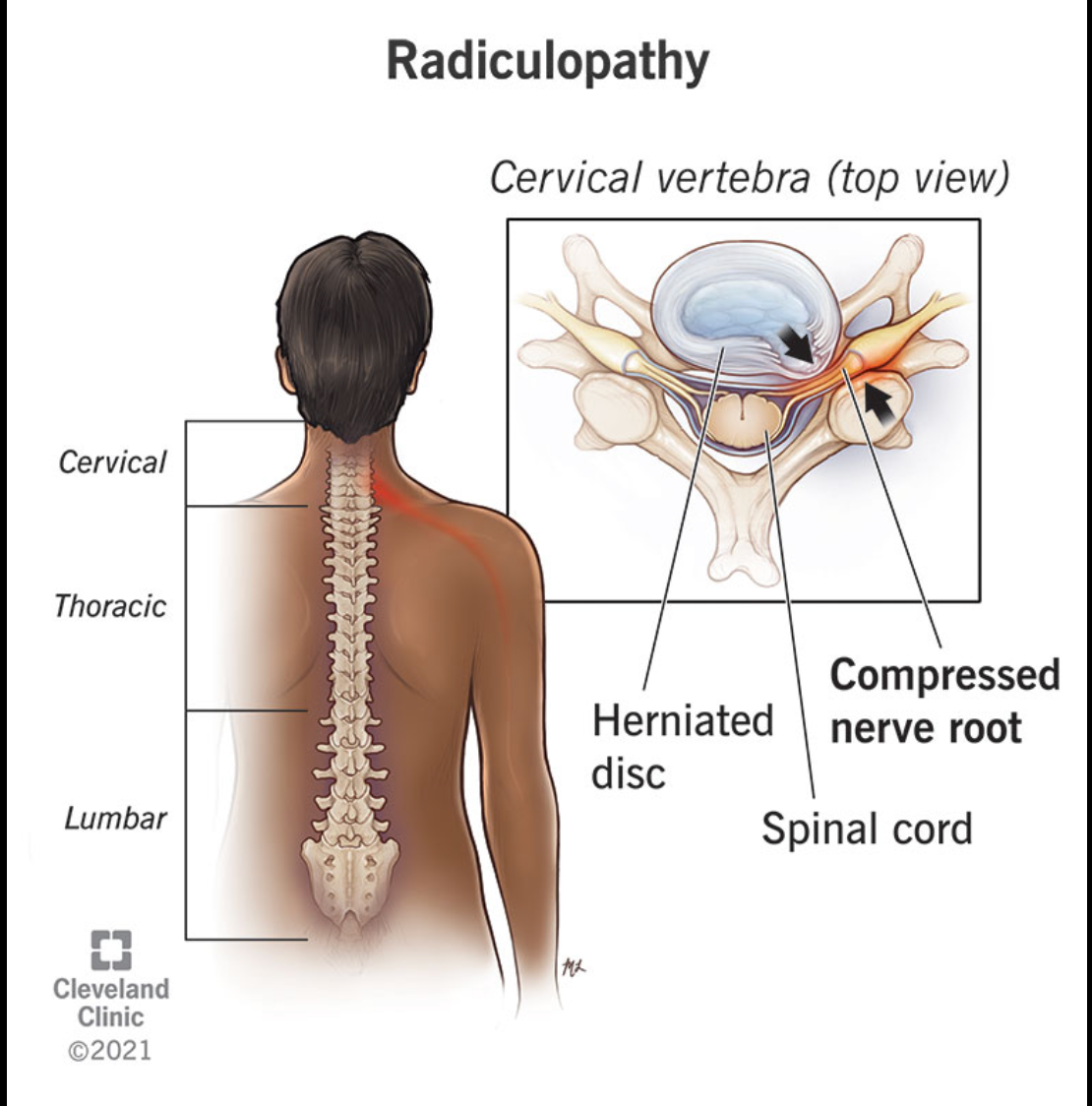
✔ Lumbar Disc Herniation
– Sciatica, leg pain, or numbness due to nerve compression.
✔ Lumbar Canal or Lateral Recess Stenosis
– Especially beneficial for elderly patients needing gentle tissue handling.
✔ Foraminal Stenosis
– Narrowing of the nerve exit pathway.
✔ Recurrent Disc Herniation
– After previous spine surgery (microdiscectomy or open surgery).
✔ Failure of Conservative Treatment
– Persistent symptoms despite medications, rest, physiotherapy, or injections.
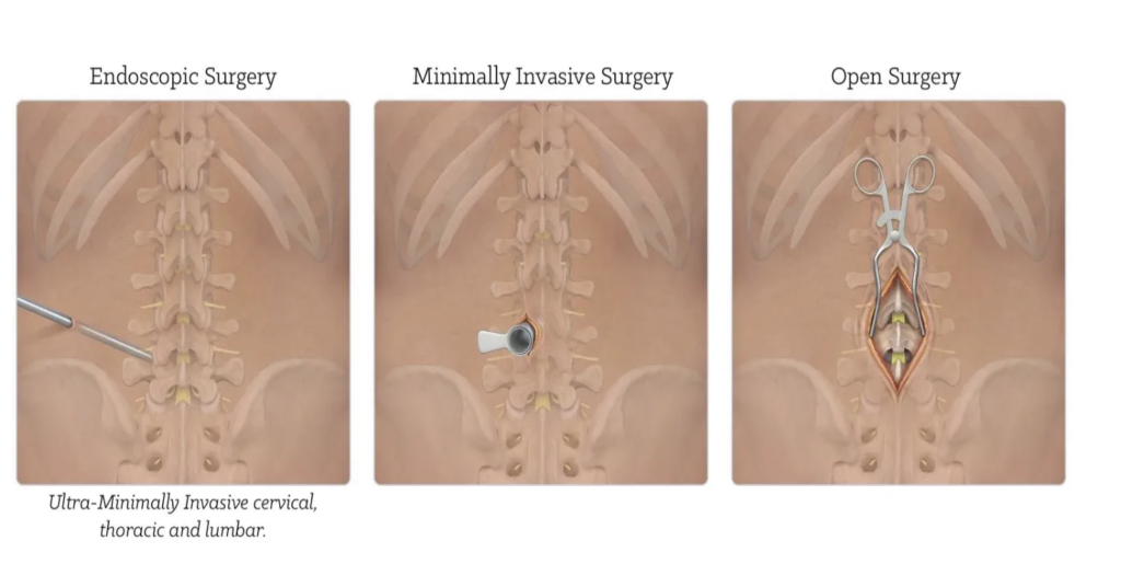
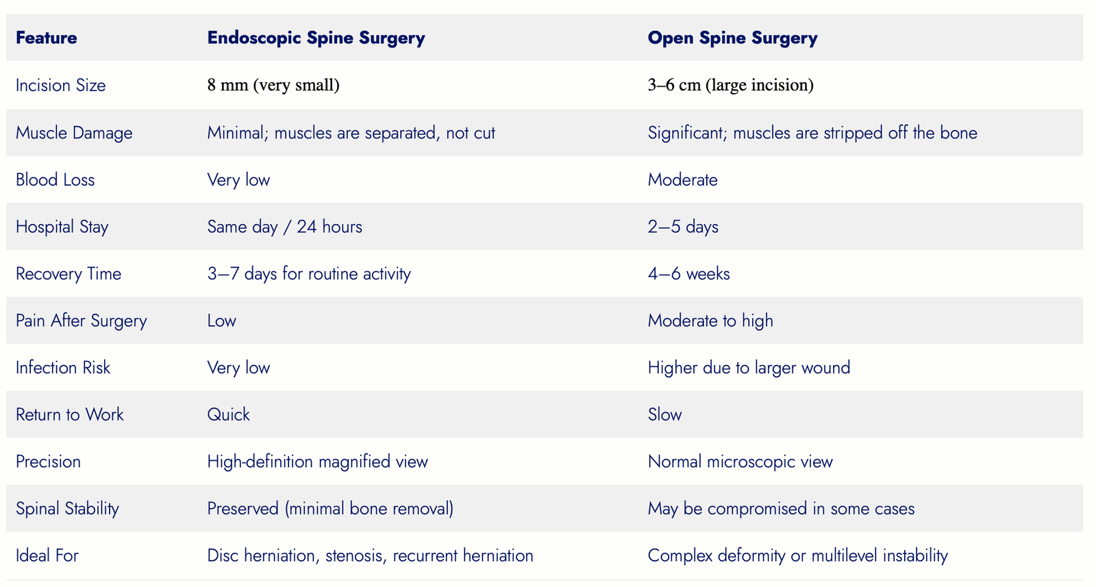
Comparison: Endoscopic vs. Open Spine Surgery
What to Expect Before Surgery
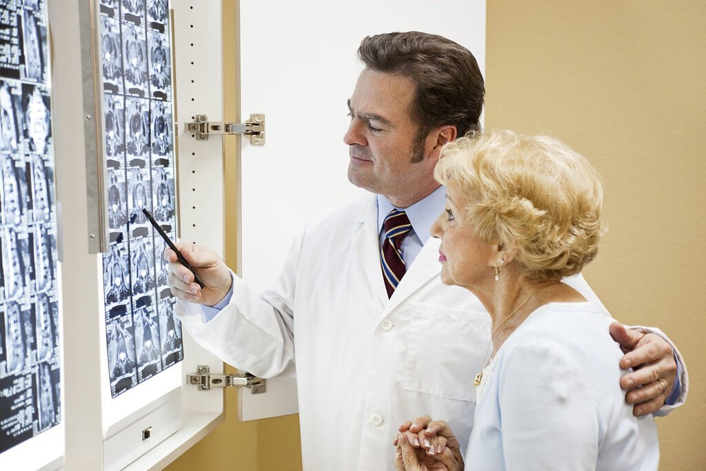
- Detailed evaluation including MRI and clinical examination
- Discussion about your symptoms, expectations, and treatment goals
- Blood tests and anesthesia assessment
- Instructions regarding fasting before surgery
- Stopping certain medications (e.g., blood thinners) if advised
- Explanation of the entire procedure and recovery plan
Our team ensures you are confident and fully prepared before the surgery
How the Surgery Works
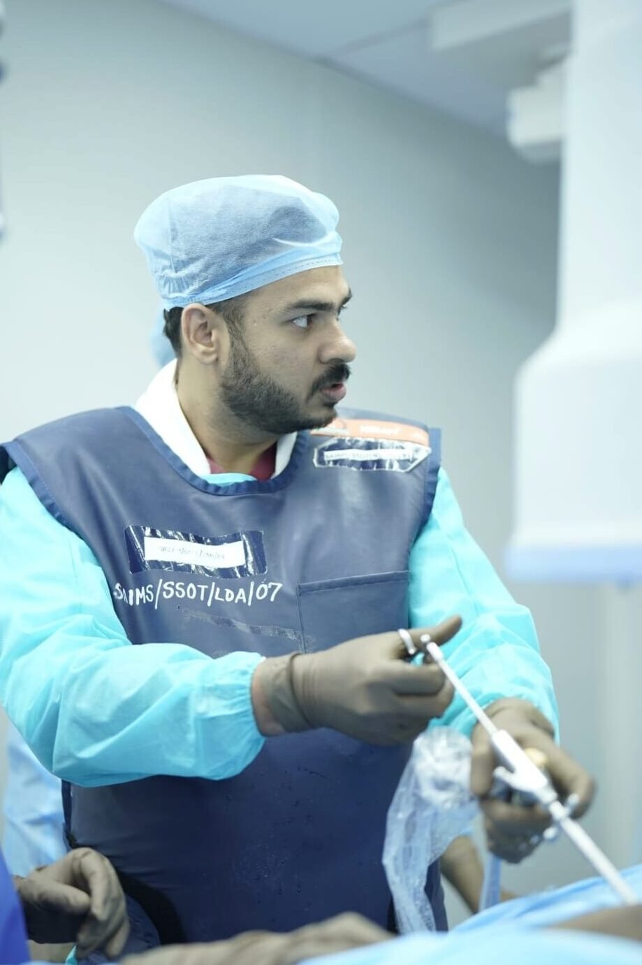
- Performed under local anesthesia with sedation or general anesthesia (case-dependent)
- A small incision (approx. 8 mm) is made in the interlaminar window
- High-definition endoscope is inserted
- Herniated disc or compressing tissue is carefully removed
- Continuous saline irrigation ensures clear visibility
- Incision is closed with one or two sutures or steri-strips
Recovery & Expectations
Patients typically experience significant relief in leg pain immediately after surgery.

- Walk within a few hours after surgery
- Same-day discharge in most cases
- Mild soreness for 1–2 days
- Resume office work within a few days
- Avoid heavy lifting for 3–4 weeks
- Follow-up evaluation after 2 weeks and 6 weeks